A cannabidiol-loaded sponge placed directly at the site of a traumatic brain injury reduced brain swelling, neuroinflammation and neurological deficits in rats, according to a newly accepted study.
The research, set to appear in Materials Today Bio, was conducted by researchers from Kunming Medical University and the First Affiliated Hospital of Kunming Medical University. The study was accepted September 30.
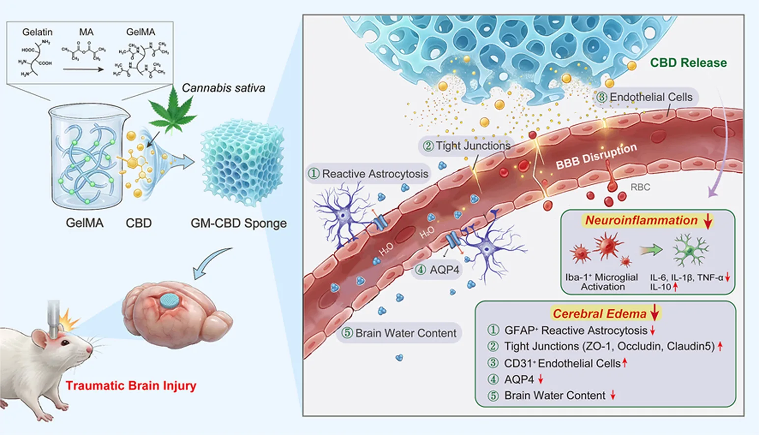
Researchers developed a biodegradable methacrylated gelatin, or GelMA, sponge containing CBD. The experimental system was designed to release CBD directly into injured brain tissue rather than relying on oral or systemic administration.
A total of 148 adult male rats were used in the study, with 146 included in the final analyses. Researchers induced traumatic brain injuries using a modified Feeney weight-drop model and compared untreated animals with groups receiving mannitol, injected CBD, a blank GelMA sponge or GelMA sponges containing different amounts of CBD.
Based on neurological and tissue assessments, researchers selected a sponge with a nominal CBD loading of 15 micrograms as the working dose.
At 48 hours after injury, rats treated with the CBD sponge had significantly lower neurological deficit scores and improvements in locomotion and exploratory behavior. Tissue examinations also found greater neuronal survival, less neuronal damage and reduced apoptosis compared with untreated animals.
Researchers found particularly notable effects on cerebral edema, one of the potentially dangerous consequences of traumatic brain injury.
The CBD sponge significantly reduced brain water content and leakage across the blood-brain barrier. Its effect on brain water content was similar to mannitol, a drug commonly used to reduce intracranial pressure, while the blank GelMA sponge produced only a modest, statistically nonsignificant change.
Researchers also found that the CBD treatment helped restore several proteins critical to blood-brain barrier integrity, including ZO-1, occludin and claudin-5.
The treatment increased expression of the endothelial marker CD31 and altered aquaporin-4, or AQP4, a water-channel protein involved in regulating fluid in the brain.
Following traumatic brain injury, AQP4 expression increased and became more closely associated with reactive astrocytes. Treatment with the CBD sponge reduced AQP4 and GFAP expression while increasing CD31 expression, changes the researchers said were consistent with reduced astrocyte activation and better preservation of vascular integrity.
The treatment also produced substantial changes in inflammatory markers.
Levels of the pro-inflammatory cytokines IL-6, IL-1β and TNF-α declined, while levels of the anti-inflammatory cytokine IL-10 increased. Microglial activation was also reduced, suggesting suppression of the acute inflammatory response caused by the injury.
One potential advantage of the approach was its ability to concentrate CBD around the injury while limiting systemic exposure.
CBD remained detectable in tissue surrounding the lesion throughout the 48-hour observation period. With the CBD sponge, cortical CBD concentrations reached 251.08 nanograms per gram at one hour and remained detectable throughout the study period.
By comparison, injected CBD produced a peak cortical concentration of 91.30 nanograms per gram.
Overall CBD exposure in the injured cortex was also substantially higher with the sponge, while CBD concentrations in blood were considerably lower.
Researchers cautioned, however, that the sponge and injected CBD treatments were not administered at equivalent doses or through equivalent routes, meaning the two approaches cannot be directly compared for efficacy.
Laboratory testing showed that the sponge provided sustained CBD release over several days.
About 22% of the CBD was released during the first two hours, increasing to about 58% after 24 hours and approximately 80% after 72 hours.
Further experiments suggested CBD may interact directly with AQP4.
Molecular modeling predicted an interaction between CBD and the protein, while surface plasmon resonance testing detected a measurable micromolar interaction between CBD and immobilized AQP4.
Researchers also conducted gene-expression analysis on brain tissue from treated rats.
The analysis found changes involving the NF-κB, PI3K-Akt and TNF signaling pathways, which are involved in inflammation, cellular survival and responses to injury.
Corresponding changes were detected in proteins associated with those pathways, along with lower TNF-α levels.
The authors cautioned that the gene-expression analysis did not include groups receiving a blank sponge or injected CBD, making it impossible to determine whether the molecular changes resulted specifically from CBD or from the localized delivery system.
Short-term safety testing found no detectable treatment-related abnormalities in major organs or measures of liver and kidney function under the conditions studied.
The researchers emphasized that the findings remain preliminary.
The study examined animals only during the first two days following traumatic brain injury, meaning it does not establish whether the CBD sponge improves long-term cognition, behavior or neurological recovery.
The researchers said future studies should examine longer-term outcomes and compare localized CBD treatments using matched doses and exposure levels.
They concluded that local administration of CBD through the GelMA sponge showed neuroprotective potential by reducing acute cerebral edema and neuroinflammation while maintaining CBD around the injured area of the brain.